The Anatomic Areas of a Long Bone
(Femur, Humerus)

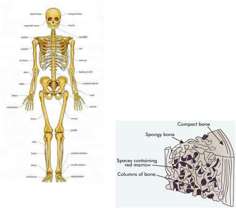
Diaphysis- shaft, compact bone
Epiphysis- End of Bone, spongy bone
Periosteum- outside covering of bone
Sharpey's Fibers- periosteum of underlying bone
Arteries- supplies the bones with nutrients
Articular Cartilage- covers external surface of epiphysis
Epiphyseal Plate- Flat plate seen in young people
Epiphyseal Line- remnant in adult bones
Medullary cavity- location of yellow bone marrow/ red bone marrow
The Creation of Bone
-The process of bone formation is called Ossification
-The hyaline cartilage model is completely covered with bone matrix by bone forming cells called osteoblasts
-By birth most hyaline cartilage models have been converted to bone except for two regions: Articular Cartilages and Epiphyseal Plates.
Bone Fractures

1- Comminuted- Bone breaks into various fragments.
2- Compression- Bone is completely crushed.
3- Depressed- Broken bone portion is pressed inward.
4- Impacted- Broken Bone ends are forced into eachother.
5- Spiral- Ragged break occurs when excessive twisting forces are applied.
6- Greenstick- Bone breaks incompletely, much in the way a twig breaks.
      |
|
